Dewachter, P., Raëth-Fries, I., Jouan-Hureaux, V., Menu, P., Vigneron, C., Longrois, D., and Mertes, P. M. 2007. A Comparison of Epinephrine Only, Arginine Vasopressin Only, and Epinephrine Followed by Arginine Vasopressin on the Survival Rate in a Rat Model of Anaphylactic Shock. Anesthesiology. 106: 977-983. PMID: 17457129
Pubmed link here!
 Figure 1: Symptoms of Anaphylaxis (From Medline Plus)
Summary:
Introduction, Materials and Methods
It has been well established in scientific literature as well as medical practice that epinephrine is the most effective in treating anaphylactic shock [1]. Anaphylactic shock is an acute systemic, severe reaction leading to systemic vasodilation (associated with a sudden drop in blood pressure) and edema of bronchial mucosa (leading to bronchoconstriction and difficulty breathing) [3]. If left untreated, this condition can result in death in a matter of minutes. Epinephrin serves to counteract these effects by directly acting on α- and β-adrenergic receptors in an agonistic fashion. Its actions on α1-adrenergic receptors increases left ventricular preload by reducing venous capacitance; this serves to increase blood pressure. Its actions on β-adrenergic receptors reverse the bronchoconstriction and increase cardiac inotropy (contractility) and chronotropy (heart rate); this serves to both increase blood pressure and relieve breathing difficulty [1, 2].
 Figure 2: Symptoms of Anaphylaxis (From Ambulance Technician Study)
Recent experimental studies have also shown that arginine vasopressin (AVP) serves to increase mean arterial pressure (MAP) values in animal models of anaphylactic shock. This increase in MAP was comparable to the increase in MAP observed when anaphylactic shock is treated with epinephrine [1]. However, even the highest AVP doses were associated with a much lower skeletal muscle oxygen pressure (PtiO2) value, a parameter previously shown in human and animal models to be correlated with survival [1].
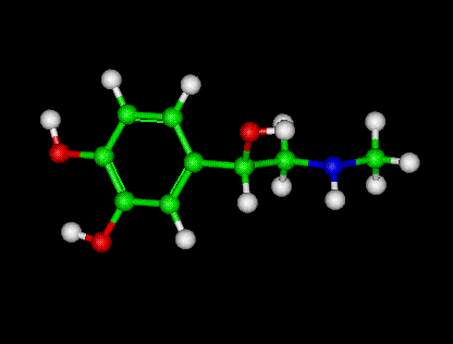
 Figure 3: Molecular model of arginine vasporessin (AVP) (From 3D Chem)
The purpose of this study are twofold: first, to compare the effects of epinephrine only and AVP only on the resolution of anaphylaxis in terms of MAP, heart rate, Ptio2 value, and survival. Second, to investigate the effects of using AVP after epinephrine treatment on anaphylaxis in terms of MAP, heart rate, PtiO2 value, and survival [1].


Results and Discussion

Graph A: Profile of Mean Arterial Pressure (MAP) before and after onset of treatment in each of the three treatment groups and the control (no treatment) group. Note that T=0 corresponds to the time in which anaphylaxis was induced. T=5 corresponds to the time at which treatment began. Note that epinephrine significantly increases MAP, as does epinephrine plus AVP but at a slower rate. AVP only causes no significant change in MAP [1] (from Dewachter et al. (2007)).

Graph B: Profile of skeletal muscle tissue oxygen partial pressure (PtiO2) after onset of treatment in each of the three treatment groups and the control (no treatment) group. Note that T=0 corresponds to the time in which anaphylaxis was induced. T=5 corresponds to the time at which treatment began. Note that epinephrine significantly increases PtiO2, as does epinephrine plus AVP but at a slower rate. AVP only does not result in any increase in PtiO2 [1] (from Dewachter et al. (2007)).
The main findings of this study were as follows: therapy with epinephrine only was associated with a high rate of survival (84%), where AVP treatment only had a 100% mortality rate; epinephrine therapy followed by continuous infusion of AVP was associated with a 100% survival rate [1]. When comparing the epinephrine only, AVP only, and epinephrine plus AVP groups, the MAP and PtiO2 profiles are very similar. The epinephrine only and epinephrine plus AVP groups were characterized by a partial restoration of MAP and a full recovery of PtiO2 associated with tachycardia and unchanged heart rate, respectively. The AVP group, on the other hand, was characterized by a decrease in MAP and PtiO2 and a low heart rate. The survival of the three groups differed as well: there was a high rate of survival in the epinephrine only group and the epinephrine plus AVP group [1]. The survival rate in the AVP only group was nonexistent; there was a 100% mortality rate. Despite the initial increase of MAP caused by AVP, it decreases more over time and is associated with a lower heart rate and a significant decrease of PtiO2 [1].
When AVP was injected as a continuous infusion after an initial dose of epinephrine, AVP restored MAP, just as epinephrine only treatment did, and was associated with an unchanged heart rate. Tachycardia was, however, observed with the epinephrine only group [1].
It is noteworthy that, when comparing the epinephrine plus AVP group to the AVP only group, there was a lower infusion of AVP in the epinephrine plus AVP group than in the AVP only group. Despite this lower infusion, the MAP and PtiO2 values of the epinephrine and AVP group were significantly higher [1]. The drug administration duration of the epinephrine plus AVP group was also less than the duration of the AVP only group. Taken together, these observations suggest that the epinephrine administration before the infusion of AVP cause a major change in the effects of the AVP that probably explains the highly improved survival rate of this group [1].
 Figure 4: Surgery being performed on a rat (From ResearchTraining.org)
The main results of this paper showed that a single treatment of epinephrine caused an increase in MAP accompanied by simultaneous increase in cardiac output and mean pulmonary capillary wedge pressure compared with the control group. A continued dosage of epinephrine produces a sustained improvement of in MAP and PtiO2 [1, 4]. It acts by increasing cardiac output and stroke work due to the effects epinephrine binding to β-adrenergic receptors. The mechanism by which AVP acts as a vasopressor is unclear. AVP, in vasoplegic shock, has been shown to restore vascular tone by at least four mechanisms. The activation of V1 receptors; the closure of ATP-sensitive K+ channels which normally produce vasodilation via cellular hyperpolarization; the modulation of nitric oxide via antagonization; and the potentiation of adrenergic and other vasoconstrictor agents [1].
Considering the results, Dewachter et al. (2007) show that epinephrine must still be considered the first line of drug treatment for anaphylaxis. They also show, however, that AVP can be used to supplement epinephrine treatment after epinephrine has been given [1].
Critique:
This paper was very clear and concise. Anesthesia-induced anaphylaxis was generated and modeled well in the Norway Rat; ultimately, this means it was an excellent model for the human system. The anaphylactic state and the mechanism of action in which it is relieved by epinephrine was explained in a clear, precise fashion. Although the mechanism through which AVP acts as a vasopressor is unclear, the authors still give several possibilities of how this mechanism may work. This was much appreciated, as it really provided a better overall picture of how AVP would fit into the anaphylaxis treatment regime. The purpose of this paper was to determine if AVP could be potentially used to supplement epinephrine as a treatment for anaphylaxis; ultimately, the authors claimed AVP could supplement epinephrine but not replace it, and the results supported this conclusion.
The figures presented in the results section were clear and well organized. They presented enough information to be helpful and show trends but not so much that they became unclear. The legends to each of the figures were excellent and easy to understand. There was, however, a problem with clarity with the table indicating the heart rate measured in the rats during anaphylaxis. It seemed rather meaningless to simply present a table of numbers. It would have been far more useful to present the heart rates in a graphical format, such as a bar graph or a line graph. This way the reader would be able to easily view trends in the heart rate, such as tachycardia or a decrease back to baseline.
It was also useful for the authors to explain the materials and methods as thoroughly as they did. Since this experiment is an animal model for a human condition, it was important to explain the function and purpose of each of the steps in a human context. For example, after anaphylaxis was induced the authors waited five minutes before administering treatment. This was meant to simulate, in a human operating room environment, the time needed for the diagnosis of anaphylaxis and the preparation of the treatment.
Overall this paper was incredibly thorough and well informed. The authors validated the continued use of epinephrine as the primary treatment for anaphylaxis and indicated the benefits of using AVP with this treatment.
Future Experiments:
Future experiments are required to validate the information obtained by the Dewachter et al. (2007) results. Although the addition of AVP to traditional epinephrine treatment of anaphylaxis may help increase survival rate, it is important to realize that these results were for rats and may not be of clinical importance in humans. The most logical step at this point would be to organize a human trial. Although it would be ethically irresponsible to induce anaphylaxis in humans in order to test the effects of AVP on epinephrine treatment, it is possible to conduct a study on patients already scheduled for surgery who may be at a high risk for anaphylaxis. If consent is obtained, these patients would agree to receive a continuous intravenous injection of AVP along with epinephrine treatment in the occurrence of anaphylaxis. Surgery requires monitoring of blood pressure, heart rate, respiratory rate, and oxygen saturation levels so no additional equipment would have to be used. After surgery, if anaphylaxis occurred, the information from these monitors can be easily downloaded and the effects of the epinephrine and AVP administration can be determined. Standards can be obtained from other surgeries where epinephrine was used on anaphylaxis to establish a baseline rate of recovery and survival rate. If the AVP in the epinephrine treatment is not successful, the statistics obtained from the surgery will closely resemble those of the standard epinephrine treatment. If the AVP enhances the epinephrine treatment, the surgery statistics will show a more rapid improvement (meaning a faster increase in blood pressure, a faster increase in oxygen saturation, and the absence of tachycardia). Only when AVP has been tested in human trials and found to be successful can we truly incorporate it into the treatment of anaphylaxis.

References:
[1] Dewachter, P., Raëth-Fries, I., Jouan-Hureaux, V., Menu, P., Vigneron, C., Longrois, D., and Mertes, P. M. (2007). A Comparison of Epinephrine Only, Arginine Vasopressin Only, and Epinephrine Followed by Arginine Vasopressin on the Survival Rate in a Rat Model of Anaphylactic Shock. Anesthesiology. 106: 977-983.
[2] Kindt, T., Goldsby, R., Osborne, B. (2007). Kuby Immunology.New York, NY: W. H. Freeman and Company.
[3] Silverthorn, D. (2007). Human Physiology: An Integrated Approach. San Francisco, USA: Benjamin Cummings.
[4] Stolk, Jon M., U’Prichard, David C., Fuxe, Kjell. (Eds.). (1988). Epinephrine in the Central Nervous System. Oxford: Oxford University Press.


